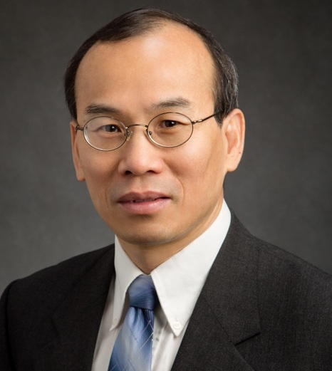
Prof. David A. Boas, PhD (USA)
David Boas, Ph.D. (Professor, Biomedical Engineering) is Director of the Neurophotonics Center at Boston University. He received his BS in Physics at Rensselear Polytechnic Institute and PhD in Physics at the University of Pennsylvania. During his academic career, he has supervised more than 50 students and post-doctoral fellows, and he has published over 300 papers that have received over 46,000 citations and an h-index of 117. He is the founding President of the Society for Functional Near Infrared Spectroscopy and founding Editor-in-Chief of the journal Neurophotonics published by SPIE. Dr. Boas was awarded the Britton Chance Award in Biomedical Optics in 2016 for his development of several novel, high-impact biomedical optical technologies in the neurosciences, as well as following through with impactful application studies, and fostering the widespread adoption of these technologies. He was elected a Fellow of AIMBE, SPIE, and OSA in 2017.
As Director of the Neurophotonics Center, he facilitates the development and application of novel optical methods to address a broad range of neuroscience questions from basic science to clinical translation. His own research efforts focus on neurovascular coupling, cerebral oxygen delivery and consumption, functional near infrared spectroscopy (fNIRS), and physiological modeling. Studies are done in rodents and humans, invasively and non-invasively, microscopically and macroscopically, providing a powerful ability to translate findings from animals to humans, and conversely to address in animals questions raised during human studies. One example of this that will tie together many of Dr. Boas’ activities is studying functional brain recovery in survivors of stroke. Human neuroimaging by fMRI and fNIRS measures hemodynamic functional recovery but it is not known if neuro-vascular coupling differs in these patients compared to healthy subjects. Animal studies will answer this question enabling more quantitative interpretation of the human neuroimaging studies.
Professor (BME, ECE)
Neurophotonics Center
Boston University College of Engineering
USA
PL1 Using light absorption and laser speckle dynamics to measure human brain function
Monday, June 16, 14:45, Lecture Hall A

Prof. Zhi-Pei Liang, PhD (USA)
Zhi-Pei Liang received his Ph.D. degree from Case Western Reserve University in 1989. He subsequently joined the University of Illinois at Urbana-Champaign (UIUC) first as a postdoctoral fellow (supervised by Nobel Laureate Paul Lauterbur) and then as a faculty member. Dr. Liang is currently the Franklin W. Woeltge Professor of Electrical and Computer Engineering; he also chairs the Computational Imaging Group in the Beckman Institute for Advanced Science and Technology. Dr. Liang’s research is in the general area of magnetic resonance imaging, signal processing and machine learning. His work has been recognized by a number of awards, including the Sylvia Sorkin Greenfield Award (Medical Physics, 1990), the Whitaker Biomedical Engineering Research Award (1991), the NSF CAREER Award (1995), the University Scholar Award (UIUC, 2001), the Isidor I. Rabi Award (ISMRM, 2009), the Otto Schmitt Award (IFMBE, 2012), IEEE-EMBC Best Paper Awards (2010, 2011, 2021), IEEE-ISBI Best Paper Awards (2010, 2015), the Technical Achievement Award (IEEE-EMBS, 2014), and the Gold Medal (ISMRM, 2022). Dr. Liang was selected as the Paul C. Lauterbur Lecturer for the 2016 ISMRM meeting and as the Savio L. Woo Distinguished Lecturer for the 2017 WACBE World Congress on Bioengineering. He is a Fellow of IEEE, ISMRM and AIMBE. He was elected to the International Academy of Medical and Biological Engineering in 2012 and to the US National Academy of Inventors in 2021. Dr. Liang served as President of the IEEE-EMBS from 2011-2012 and received its Distinguished Service Award in 2015. He has been serving as Chair-elect (2022-2024) and Chair (2025-2027) of the International Academy of Medical and Biological Engineering.
Franklin W. Woeltge Professor
Department of Electrical and Computer Engineering, and
Beckman Institute for Advanced Science and Technology
University of Illinois at Urbana-Champaign
USA
PL2 AI-powered MRI: Towards an Omni Imaging Technology for Brain Mapping
Monday, June 16, 15:35, Lecture Hall A
The ongoing paradigm shift in healthcare towards personalized and precision medicine is posing a critical need for noninvasive imaging technology that can provide quantitative tissue and molecular information. Magnetic resonance signals from biological systems contain information from multiple molecules and multiple physical/biological processes (e.g., T1 relaxation, T2 relation, diffusion, perfusion, etc.). So, magnetic resonance imaging (MRI) is inherently a high-dimensional imaging technology that can acquire structural, functional and molecular information simultaneously. In practice, due to the curse of dimensionality, MRI experiments are often done in a low-dimensional setting to acquire biomarkers one at a time. Such a “divide-and-conquer” approach not only reduces data acquisition efficiency but also makes it difficult to obtain molecular information in high resolution. By synergistically integrating machine learning with sparse sampling, constrained image reconstruction and quantum simulation, we have successfully demonstrated ultrafast high-dimensional imaging of the brain. This talk will give an overview of this unprecedented omni imaging technology and show some exciting experimental results of brain function and diseases.

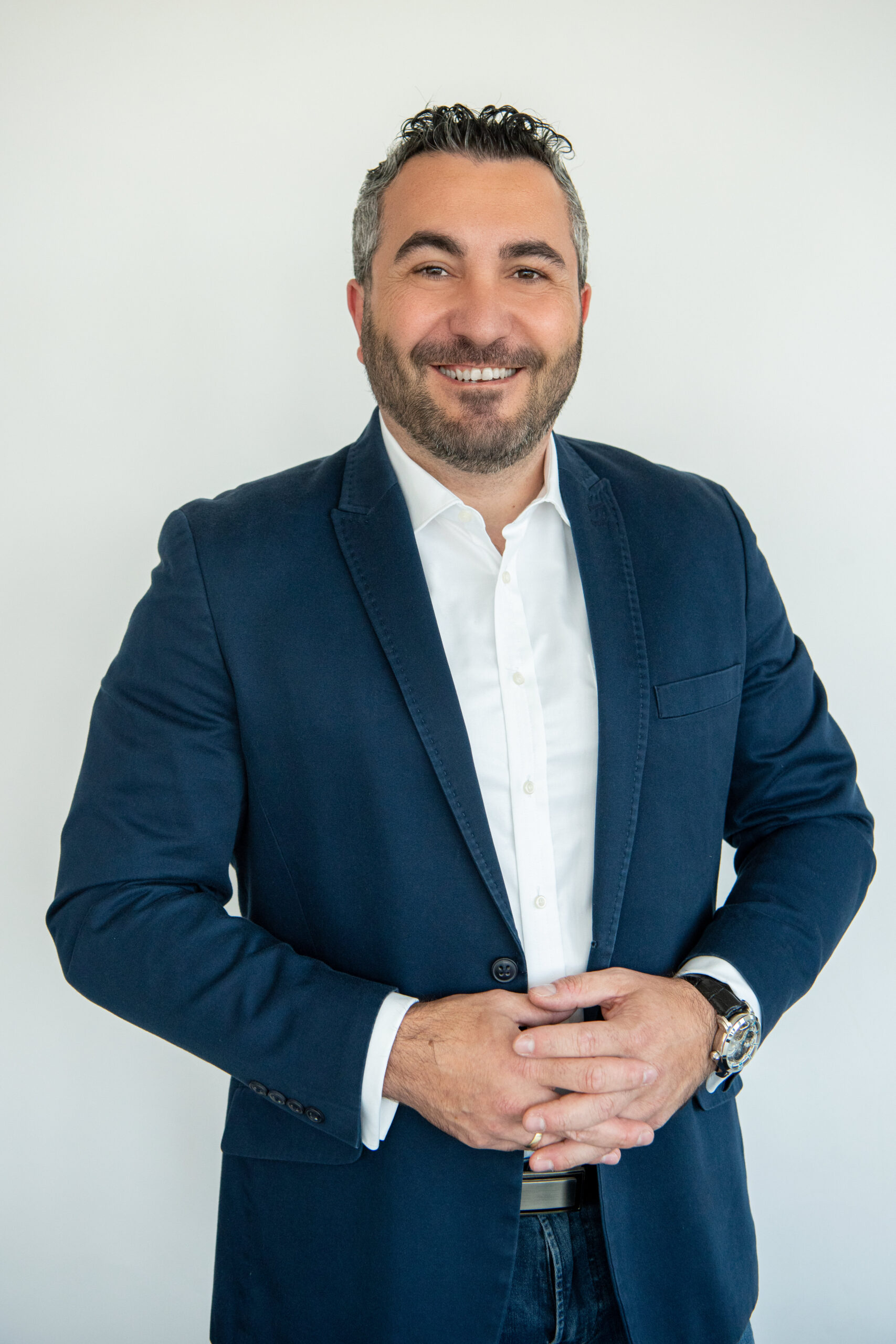
Dr. Bassil Akra (Germany)
Dr. Bassil Akra is CEO and President of AKRA TEAM in Germany (GmbH) and the USA (Inc), a globally acting consultancy company. Dr. Akra was the Vice President of Strategic Business Development at the Global Medical Health Services of TÜV SÜD Product Service GmbH. He has vast experience in leadership, business management, research, development, quality management, and regulatory approval of medical devices, combination devices, and ATMP Products. Dr. Akra played an essential role during the implementation of the medical device regulation in Europe. He was also involved in the drafting of several European guidance documents (e.g. MEDDEV, MDCG, etc.) and International Standards. He spent the last years of his career at TÜV SÜD training and educating various stakeholders on EU Legislations. Dr. Akra is also member of the Board of the EU Association TEAM PRRC. He is also member of the MDR EY Survey Advisory Group.
CEO, AKRA TEAM GmbH
PL3 European Medical Device CE Mark – Current challenges and future opportunities
Tuesday, June 16, 14:45, Lecture Hall A

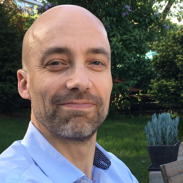
Prof. Lauri Parkkonen, PhD (Finland)
The main research area of Parkkonen is non-invasive neuroimaging, mainly instrumentation and data analysis methods related to MEG as well as human neuroscience of sensory modalities and cognition. He has contributed to MEG instrumentation and signal processing methods that are currently in active use in over 100 laboratories world-wide. Currently he pursues the use of optically-pumped magnetometers for MEG, exploits real-time feedback of measurements of brain activity to study cognition and plasticity, both for basic neuroscientific research and for clinical applications. He is also coordinating a national consortium to develop a biobank for functional brain-imaging data.
Parkkonen has also contributed to ultra-low-field MRI research and to invasive monitoring of local cortical activity. In cognitive neuroscience, Parkkonen has shed light on the neural mechanisms of sensory processing, particularly conscious visual perception. Recently he has pioneered the emerging field of social neuroscience to uncover brain mechanisms supporting social interaction by using hyperscanning i.e. simultaneous recording of the brain activity of the interacting participants.
Aalto Brain Centre
Department of Neuroscience and Biomedical Engineering,
Aalto University School of Science
Finland
PL4 High-resolution magnetoencephalography: a new window to human brain function?
Wednesday, June 18, 9:00, Lecture Hall A
